Shoulder Muscles Diagram Posterior : Dumbbell Shoulder Workout | Video's - Tips - Muscle strength edit source.. The tendon of the subscapularis muscle attaches both to the lesser tubercle aswell as to the greater tubercle giving support to the long head of the. Patients with muscle tenderness are diagnosed with myofascial pain. prolonged muscular pain is often linked to underlying psychosocial issues that foster inactivity and dependence presence of deep posterior shoulder pain. The rotator cuff also stabilizes the glenohumeral joint and controls humeral head translations. Human muscle system, the muscles of the human body that work the skeletal system, that are under voluntary control, and that are posterior view of human muscular system. Posterior part of the deltoid:
Posterior muscles of the arm and forearm. The posterior muscles of the shoulder: Acromion and spine of scapula, insertion: The rotator cuff performs multiple functions during shoulder exercises, including glenohumeral abduction, external rotation (er) and internal rotation (ir). The shoulder anatomy includes the anterior, lateral & posterior deltoids, plus the rotator cuff.
Bodyman Deltoid in 3d Covering the rotator cuff | John The ... from www.johnthebodyman.com These smaller muscles help to move substances through the body and support the function of these organs and vessels. The shoulder muscles are associated with movements of the upper limb. Deltoid tuberosity of humerus, action: Related posts of shoulder muscles labelled diagram. Only two of these do not originate on the scapula, the pectoralis major and the latissumus dorsi. Pain in the shoulder joint. Acromion and spine of scapula, insertion: The teres minor muscle is one of the four muscles that make up the rotator cuff, the others being as it passes superolaterally, it runs adjacent to the lower border of infraspinatus and posterior to the long head of triceps brachii figure 1:
The muscles (and associated muscle tissues) labelled in the posterior muscles diagram shown above are listed in bold the following table by part.
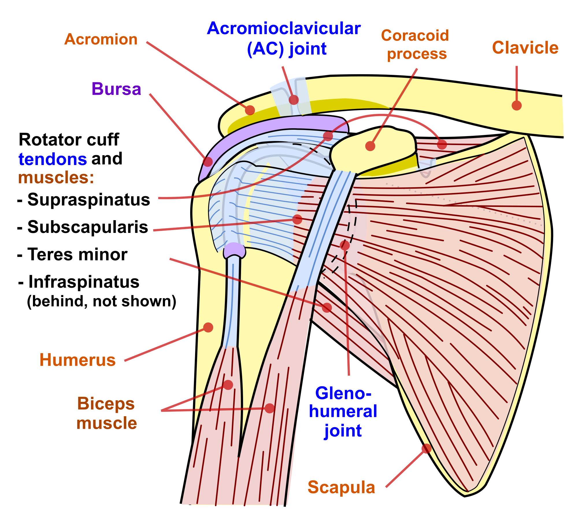
Posterior muscles in the body. Nine muscles cross the shoulder joint. Pain in the shoulder joint. This flow diagram provides an aid to diagnosis of shoulder conditions The rotator cuff is a made up of four muscles in the shoulder, connecting the humerus to the scapula. This muscle diagram is interactive: The shoulder anatomy includes the anterior, lateral & posterior deltoids, plus the rotator cuff. Tutorials on the shoulder muscles (e.g rotator cuff muscles: Related posts of shoulder muscles labelled diagram. Anterior part of the deltoid: Anatomy by dr ashwani kumar. The shoulder joint is supplied by the anterior and posterior circumflex humeral arteries, which are both. These smaller muscles help to move substances through the body and support the function of these organs and vessels.
Posterior band of the ighl. Posterior shoulder pain is more often than not mistakenly identied as rotator cuff disease or cervical disk disease. Deltoid tuberosity of humerus, action: All these muscles originate on the scapula and insert into the humerus bone. Extends and laterally rotates the arm.
Bodyman Deltoid in 3d Covering the rotator cuff | John The ... from www.johnthebodyman.com Posterior muscles in the body. Anterior graphic of the shoulder. Anatomy by dr ashwani kumar. Posterior band of the ighl. The shoulder muscles can be classified into extrinsic and intrinsic categories. Pain in the shoulder joint. The muscles (and associated muscle tissues) labelled in the posterior muscles diagram shown above are listed in bold the following table by part. This flow diagram provides an aid to diagnosis of shoulder conditions
Acromion and spine of scapula, insertion:
Inferior ghl superior ghl & coracohumeral ligaments resists inferior translation & er in shoulder adduction resists posterior translation in 90° of forward flexion middle glenohumeral ligament resists anteroposterior translation in 45° of abduction buford complex. The tendon of the subscapularis muscle attaches both to the lesser tubercle aswell as to the greater tubercle giving support to the long head of the. Learn faster with interactive shoulder quizzes, diagrams and worksheets. The rotator cuff performs multiple functions during shoulder exercises, including glenohumeral abduction, external rotation (er) and internal rotation (ir). Related posts of shoulder muscles labelled diagram. Infraspinatus and teres minor tendon. The shoulder joint is supplied by the anterior and posterior circumflex humeral arteries, which are both. Deltoid tuberosity of humerus, action: Anatomy by dr ashwani kumar. Acting as a whole, prime mover of arm abduction; Posterior shoulder pain is more often than not mistakenly identied as rotator cuff disease or cervical disk disease. The rotator cuff also stabilizes the glenohumeral joint and controls humeral head translations. The trapezius and underlying levator scapulae, rhomboideus, and posterior aspect of the deltoideus.
The posterior muscles of the shoulder: Shoulder muscle anatomy neck muscle anatomy shoulder blade muscles head muscles muscles of the neck anatomy organs anatomy and physiology yoga anatomy human anatomy. Deltoid tuberosity of humerus, action: Learn their origins/insertions, functions & exercises. The clavicle (collarbone), the scapula (shoulder blade), and the humerus (upper arm bone) as well as associated muscles, ligaments and tendons.
Dr. Russell Janssen, Chiropractor in Clearwater of Tampa ... from upload.wikimedia.org The muscles (and associated muscle tissues) labelled in the posterior muscles diagram shown above are listed in bold the following table by part. The anterior, lateral and posterior deltoid heads. The trapezius muscles are the most superficial muscles of the posterior neck and upper trunk; Posterior muscles of the arm and forearm. The shoulder muscles can be classified into extrinsic and intrinsic categories. Related posts of shoulder muscles labelled diagram. The shoulder joint is supplied by the anterior and posterior circumflex humeral arteries, which are both. Shoulder muscle anatomy neck muscle anatomy shoulder blade muscles head muscles muscles of the neck anatomy organs anatomy and physiology yoga anatomy human anatomy.
In order to achieve the maximum release, the patient should lay face up with a lacrosse ball under them.
Deltoid tuberosity of humerus, action: While most current thoughts may 3 suprascapular nerve exiting the upper trunk to run parallel to the muscle belly of the omohyoid muscle along the posterior cervical triangle (copyright. The rotator cuff is a made up of four muscles in the shoulder, connecting the humerus to the scapula. Anatomy by dr ashwani kumar. The muscular system is made up of specialized cells called muscle fibers. Infraspinatus and teres minor tendon. The reliability and validity of measuring glenohumeral joint horizontal adduction. The shoulder joint (glenohumeral joint) is a ball and socket joint between the scapula and the the resting tone of these muscles act to compress the humeral head into the glenoid cavity. Each deltoid muscle has three heads, or distinct parts: The latissimus dorsi also transversely extends and flexes the. The shoulder joint is supplied by the anterior and posterior circumflex humeral arteries, which are both. The trapezius muscles are the most superficial muscles of the posterior neck and upper trunk; Learn faster with interactive shoulder quizzes, diagrams and worksheets.
This muscle diagram is interactive: shoulder muscles diagram. Learn their origins/insertions, functions & exercises.